 Functional magnetic resonance imaging (fMRI), a method for safely measuring brain activity, has been around for about 15 years. Within the last 10 of those years a revolutionary, if mysterious, method has been developing using the technology. This method, resting state functional connectivity (rs-fcMRI), has recently gained popularity for its putative ability to measure how brain regions interact innately (outside of any particular task context).
Being able to measuring innate functional brain connectivity would allow us to know if a set of regions active during a particular task is, in fact, well connected enough generally to be considered a network. We could then predict what brain regions are likely to be active together in the future. This could, in turn, motivate us to look deeper at the nature of each brain region and how it contributes to the neuronal networks underlying our behavior.
Rs-fcMRI uses correlations of very slow fluctuations in fMRI signals (< 0.1 Hz) when participants are at rest to determine how regions are connected. The origin of these slow fluctuations has been unclear.
Some have argued that the thoughts and day dreams of participants “at rest” may explain the strong correlations typically found between brain regions. Recently, Vincent et al., 2007 sought to address this possibility using fMRI with anesthetized monkeys.
The idea is that if unconscious monkey brains show low-frequency correlated activity across known brain networks, then such findings in humans at conscious rest are likely not due to spurious thoughts, but something more innate.
Why monkeys? Besides being more ethically acceptable to anesthetize them, there are several extremely well-studied brain networks in macaque monkeys. If, as expected, these networks show highly correlated fluctuations without consciousness, rs-fcMRI is likely valid as a method for determining innate brain connectivity.
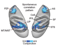
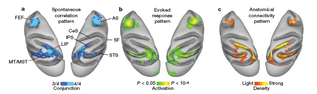
The figure presented below shows two major nodes of the monkey eye-movement control system. Part A shows the parts correlated while the monkeys were unconscious (rs-fcMRI). Part B shows the parts correlated during an eye-movement task, while part C shows the parts that are anatomically connected with brain area LIP (the lower region, within the parietal lobe).
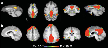
The second figure (below) shows the similarity of rs-fcMRI connectivity in monkeys (top) and humans (bottom). This is a network that is typically more active when humans are resting than when they are actively performing a task.
These results strongly support the interpretation of rs-fcMRI as measuring innate functional brain connectivity. Further, based on part C of the first figure above, rs-fcMRI may reflect anatomical connectivity as well.
What could be the origin of the correlated slow fluctuations underlying rs-fcMRI?
As pointed out by Vincent et al., 2007:
Most of the brain’s energy consumption is devoted to ongoing metabolic activity not clearly associated with any particular stimulus or behavior.
This ongoing metabolic activity may be partially driven by random neural burstings throughout the brain.
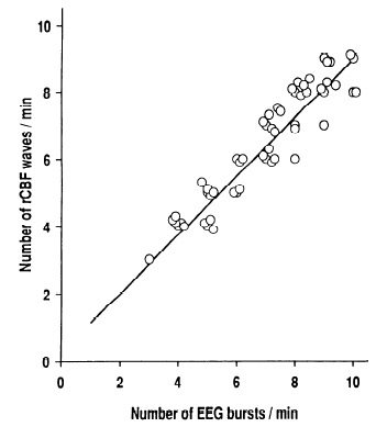
Supporting this view, a study by Golanov et al., 1994 recorded regional blood flow and electrical brain activity simultaneously in anesthetized rats. They found an extremely high correlation (88%, r=.939) between infrequent neural bursts and changes in regional blood flow (related to the fMRI signal). This relationship is shown in the figure below.
For whatever reason, it appears that brain regions burst periodically (about every 10 seconds (0.1 Hz) or so). Such bursts are necessarily sent over anatomical connections to other networked brain regions (there is no ‘shut-off switch’ for anatomical connections). These bursts should, therefore, be correlated between well-connected brain regions.
Some anatomically connected regions may not be well-correlated with rs-fcMRI, however.
Why? Simply because the strength of synaptic weights (likely governed by Hebbian learning) also determine the amount of interaction between brain regions. This predicts that the best-connected brain regions according to rs-fcMRI must be anatomically connected (directly or indirectly) and used together often during stimulus processing and behavior.
Much about the signals underlying rs-fcMRI remains to be understood. These findings give sufficient grounding to the method, however, to allow for important new discoveries about innate functional brain connectivity based on rs-fcMRI. [For instance, see recent work demonstrating an innately connected network for executive control (Cole & Schneider, 2007).]
-MWCole

References:
Vincent, J.L., Patel, G.H., Fox, M.D., Snyder, A.Z., Baker, J.T., Van Essen, D.C., Zempel, J.M., Snyder, L.H., Corbetta, M., Raichle, M.E. (2007). Intrinsic functional architecture in the anaesthetized monkey brain. Nature, 447(7140), 83-86. DOI: 10.1038/nature05758
Golanov, E.V., Yamamoto , S., Reis , D.J. (1994). Spontaneous waves of cerebral blood flow associated with a pattern of electrocortical activity. American Journal of Physiology- Regulatory, 266, 204-214.
COLE, M., SCHNEIDER, W. (2007). The cognitive control network: Integrated cortical regions with dissociable functions. NeuroImage, 37(1), 343-360. DOI: 10.1016/j.neuroimage.2007.03.071
you should have given the description of functional and anatomical parts of brain with their respective diagrams