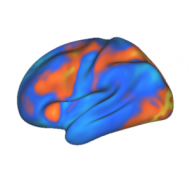
In the dark confines behind our eyes lies flesh full of mysterious patterns, constituting our hopes, desires, knowledge, and everything else fundamental to who we are. Since at least the time of Hippocrates we have wondered about the nature of this flesh and its functions. Finally, after thousands of years of wondering we are now able to observe the mysterious patterns of the living brain, with the help of neuroimaging.
First, electroencephalography (EEG) showed us that these brain patterns have some relation in time to our behaviors. EEG showed us when things happen in the brain. More recent technologies such as functional magnetic resonance imaging (fMRI) then showed us where things happen in the brain.
It has been suggested that true insights into these brain patterns will arise when we can understand the patterns’ complex spatio-temporal nature. Thus, only with sufficient spatial and temporal resolution will we be able to decipher the mechanisms behind the brain patterns, and as a result the mechanisms behind ourselves.
Magnetoencephalography (MEG) may help to provide such insight. This method uses superconducting sensors to detect subtle changes in the magnetic fields surrounding the head. These changes reflect the patterns of neural activity as they occur in the brain. Unlike fMRI (and similar methods), MEG can measure neural activity at a very high temporal resolution (>1 kHz). In this respect it is similar to EEG. However, unlike EEG, MEG patterns are not distorted by the skull and scalp, thus providing an unprecedented level of spatio-temporal resolution for observing the neural activity underlying our selves.
Despite being around for several decades, new advances in the technology are providing unprecedented abilities to observe brain activity. Of course, the method is not perfect by any means. As always, it is a method complimentary to others, and should be used in conjunction with other noninvasive (and the occasionally invasive, where appropriate) neuroimaging methods.
MEG relies on something called a superconducting quantum interference device (SQUID). Many of these SQUIDs are built into a helmet, which is cooled with liquid helium and placed around the head. Extremely small magnetic fields created by neural activity can then be detected with these SQUIDs and recorded to a computer for later analysis.
I recently got back from a trip to Finland, where I learned a great deal about MEG. I’m planning to use the method to observe the flow of information among brain regions during cognitive control tasks involving decision making, learning, and memory. I’m sure news of my work in this area will eventually make it onto this website.
-MC
I’m glad some are keeping a cool head. I suppose it might be nice to do a little TMS too, but them’s just party tricks.
@mt
Party tricks? Sure, until scientific hypotheses are clearly tested with these methods. The more methods applied to test a hypothesis the more likely it will be shown plausible or disproven (which is, of course, the whole point of science). It looks as though understanding neuronal network dynamics (the area my hypotheses are in) is going to require high temporal and spatial resolution, which will require a few of these ‘party tricks’ to perform convincing experiments.
You seem to think I was dissing MEG. It was TMS I was calling a party trick, which I was doing facetiously, except of course that TMS really is good for a few stupid tricks that would be a hit at a party (“watch me make Mary kick Joe!”). My premise was that choosing MEG means you won’t be doing TMS, since they’re both magnetic and seemed bound to interfere. Also Massimini et al referred to something like a “magnet-friendly” 60-sensor hi-res EEG helmet for the sleep study in which they’re 3D recording and pinging heads by TMS at the same time. Anyway, I think MEG is plenty cool.
@mt
I actually thought you were claiming that all of neuroimaging is a collection of ‘party tricks’. I don’t think TMS is any more of a ‘party trick’ than the other methods. In fact, it’s somewhat more powerful because it can allow for true causal inferences. Rather than saying brain region A is correlated with behavior X (what neuroimaging does), TMS can be used to say that (in the best case) brain region A is essential for behavior X. This is possible if TMSing brain region A stops or degrades the performance of the behavior. There are several papers showing these kinds of effects.
The main problem I see with TMS now is that it’s not going to just disrupt the region being TMSed. Most researchers using the method seem to ignore the fact that whole networks are going to be stimulated, since any given region in the cortex is connected to others. There is some hope, though. Concurrent neuroimaging may help with the problem, since we could see what’s being stimulated and make our inferences based on that. Alternatively, we could figure out with neuroimaging how regions are connected, then infer that TMSing region A likely also affects connected region D, which may be part of the story in disrupting behavior X.
Ultimately, all methods are ‘party tricks’ if not used correctly. Or, of course, if used to impress others at a party…
One could say that anything but clinical application is a party trick. I think a most useful outcome from MEG would be to intersect with the EEG biofeedback world which can treat a wide variety of conditions with remarkable success.
MEG is too expensive for neurofeedback, but an EEG biofeedback, or an HEG hemoencephalography system can be just a few thousand dollars. There are many opportunities for MEG to settle some controversies with this community of clinicians and researchers who are, on a daily basis, reducing and even eliminating symptoms of autism, ADHD, migraine, seizures, etc. etc.
@Gary Ames
I strongly disagree that anything but clinical application is a party trick. Basic science is extremely important for understanding the world around us, which is valuable in itself.
Additional value can typically be derived from basic research because of its usefulness to subsequent clinical research. After all, we must understand how the normal brain works before we can recognize the brain deficits present in abnormal brains.