 Most researchers in neuroscience use animal models.
Though most neuroscientists are interested in understanding the human brain, they can use more invasive techniques with animal brains. In exchange for these invasive abilities they must assume that other animals are similar enough to humans that they can actually learn something about humans in the process.
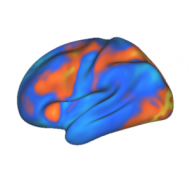
Functional magnetic resonance imaging (fMRI) is a non-invasive technique for measuring changes in local blood flow (which are significantly correlated with changes in neural activity) in the brain. fMRI measures what is called the blood oxygen level-dependent (BOLD) signal. Because it is non-invasive it can be used with human subjects.
Researchers like ourselves recognize the value of animal research, especially when the behavior being investigated is similar between the studied species and humans.
However, there is at least one fundamental cognitive difference between humans and all other animals, and likely many more given the dominant position of our species.
For researchers like ourselves it is much more interesting to learn something about the human brain (the item of interest) rather than, say, the rat brain.
Why do some neuroscientists think that using fMRI to study the neural basis of cognition in humans is of little value?
Many have heard that there are issues with fMRI as a technique. There are (like any technique), but not as many as most believe.
Here are some common misconceptions about fMRI:
- "Findings in fMRI are usually not replicated"
In fact, findings are constantly being replicated in the field. Replications are most often accomplished with slight variants on the original (e.g., 3 item working memory load rather than 2), which often increases the generality of the finding. Also, reliability of the fMRI signal is well established (see Swallow, Braver et al., 2003). Early fMRI studies had issues with replication, mostly due to poor statistical methods [see this page for some examples]. For instance, statistical thresholds were often set arbitrarily, leading to a multiple comparisons problem. The increasing use of meta-analysis in the field has shown the high extent to which findings are reproduced across studies (see this issue of Human Brain Mapping). - "Working with humans is 'messy' behaviorally"
While humans may tend to let their mind wander more than animals (an unproven claim), such mind wandering should simply add noise to the signal since it should be a pseudo-random process (all subjects don't think about the same things across task repetitions). - "fMRI is simply a new phrenology"
Phrenology was not science, while studies using fMRI can be. Findings were not replicated across individuals or studies with phrenology, which made any reported finding non-scientific. As covered above, fMRI findings are replicated on a regular basis. Also, unlike phrenology, fMRI is used for more than just functional localization (see below). - "fMRI can only measure localization of function"
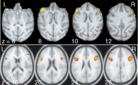
Early fMRI studies all attempted to localize functions to single regions. New conceptual frameworks (i.e., hypotheses regarding networks rather than individual regions) have changed this. Research on connectivity patterns across the brain using fMRI emphasizes distribution of processing rather than simple localization (see below). Also, new pattern recognition techniques are allowing for detection of distributed representations across the brain (see Norman et al., 2006). - "fMRI cannot be used to look at inter-regional connectivity"
New techniques have emerged to allow for inter-regional connectivity estimates using fMRI. This expands the use of fMRI beyond functional localization. The picture of brain organization that is emerging with fMRI is that of functionally specialized regions interacting to form specialized networks for complex adaptive functions. Techniques that are being used to look at connectivity include granger causality (see Goebel et al., 2003), dynamic causal modeling (see Friston et al., 2003), and resting state functional connectivity (see Cordes et al., 2000), among others (see Stephan, 2004 for review). - "No one understands what fMRI is measuring"
Several recent non-human primate studies have shown significant correlations between fMRI signal and local electrical activity (e.g., Logothetis et al., 2003). A study in rats found an extremely high correlation (R=0.94) between spontaneous electrical activity and changes in regional cerebral blood flow (Golanov et al., 1994), which is measured by fMRI. This review article (Nakahara et al., 2007) outlines a number of fMRI studies performed with both humans and non-human primates, along with single-unit recording in the non-human primates, which shows how fMRI relates to findings with more invasive methods. Single unit recording has also been done in human epileptic patients along with fMRI, showing that the signals are highly correlated (R=0.75; Mukamel et al., 2005). Consensus has emerged that fMRI is primarily a measure of changes in metabolic demand (reflected in changes in blood oxygenation) due to local changes in neural activity. - "fMRI only measures inputs to a region"
Since fMRI measures changes in metabolic demand, and most metabolic demand occurs at the synapse, it would seem that the BOLD signal only reflects inputs to a region (not outputs). However, there are typically millions of intra-regional synapses also at work within a region, such that intra-regional metabolic demand will also cause BOLD activity. Thus, most fMRI findings likely reflect intra-regional activity (not just regional inputs). Still, it seems plausible that a region may receive input (thus causing BOLD signal) without ever reaching a high enough activity level to cause any output. This allows for the possibility that fMRI can show regional activity without it ever having any effect either inside or outside that region. This is very unlikely, however, since any input large enough to cause synchronous metabolic demands across the millions of synaptic inputs to a region (a requirement for an observable fMRI response) is likely to cause massive intra-regional neural activity as well as corresponding regional output. Of course, newly developed connectivity techniques (described above) could test this by showing if a region is outputting signals to another region. - "fMRI isn't informative because it cannot distinguish between excitatory and inhibitory activity"
It's true that fMRI cannot distinguish between excitatory and inhibitory activity. However, prominent theories of cortical organization posit a canonical circuit (a cortical column) of inhibitory and excitatory neurons. Metabolic demand within these columns can therefore inform us of processing that can be further investigated using more invasive techniques. Also, a prominent theme in neuroscience involves localization of function, and fMRI can localize large-scale circuits for further investigation with other techniques that can shed light on the balance of excitatory and inhibitory connections within these circuits.
This last point suggests a segregation of function between human and animal research: Human research should involve understanding large-scale neural dynamics while animal research should involve small-scale mechanistic underpinnings of these large-scale dynamics.
Computational and mathematical modeling might be a good means for bridging these two approaches within a single framework.
fMRI is not the perfect neuroscientific method. Neither is any other method used to date. The approach the best scientists take is to ask questions, form hypotheses, and use whatever methods are available to best test those hypotheses.
This post outlined some common misconceptions that may unnecessarily limit the use of fMRI for testing scientific hypotheses.
Feel free to comment on other strengths and weaknesses of fMRI, and how other methods might be useful in conjunction with this method.
-MC & PL